Case Study18th January 2024
Revolutionizing Pediatric Cardiac Care at Southampton Hospital
Reducing surgical time - glenoid cavity pathology
Case Study
A 3D printed model was used to improve the understanding of the extent of a developmental glenoid pathology.
Case
An elderly female patient was admitted to the Ulster Hospital with extensive pain and restricted movement in her left shoulder. The patient had previously undergone bone resurfacing of the humeral head and erosion of the glenoid cavity had started because of surrounding muscular anatomy failing. Both metallic artifact and complex anatomical arrangement making image interpretation difficult.
Solution
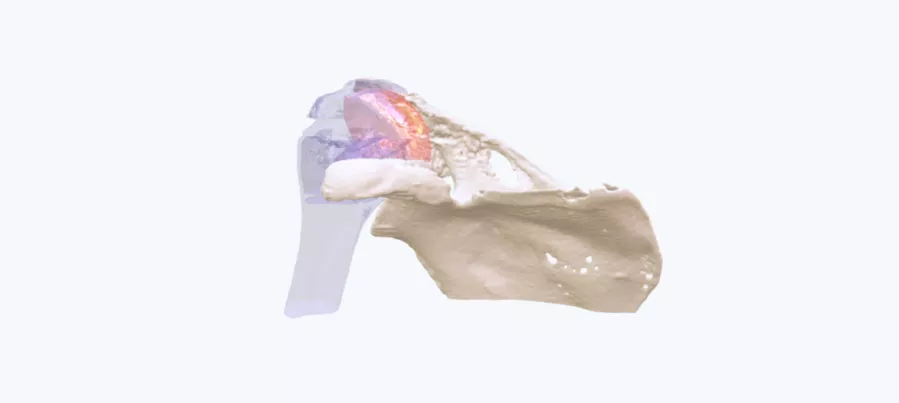
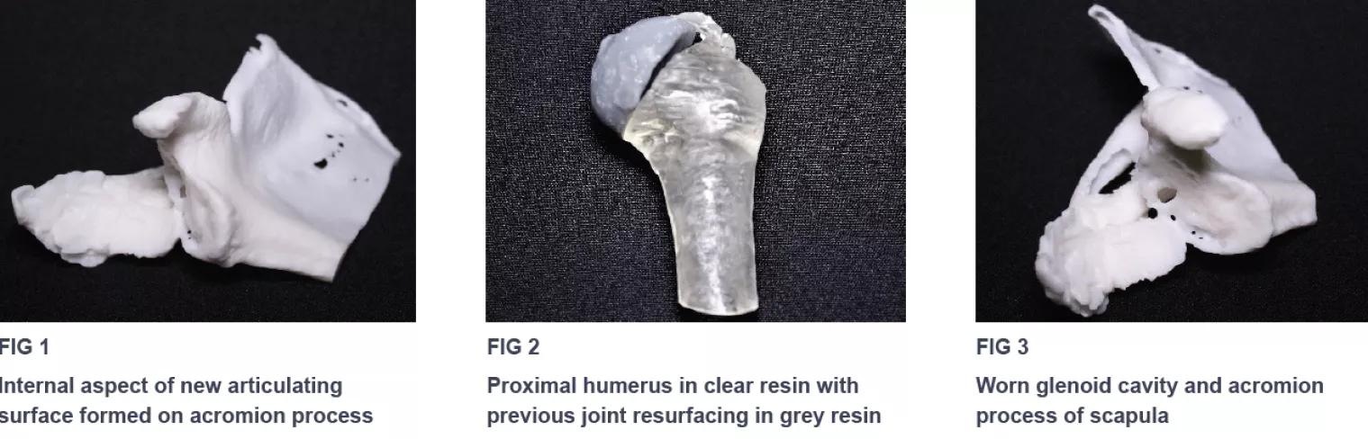
The surgeon was provided with a 1:1 scale 3D printed model of the shoulder girdle including the metallic shoulder resurfacing component (including proximal aspects of the humerus and superior portion of the scapula (Fig 1, 2 & 3).
"I was provided with a 3D model of a complex shoulder pathology which allowed me to see exactly where the glenoid had worn over time due to the resurfaced humerus and where the new articulating surface had formed superiorly to this. The models allowed me to plan with a much greater level of accuracy and the extra information provided avoided any surprised during surgery.”
- Mr. A. Adair, Consultant Orthopedic Surgeon, Ulster Hospital, Northern Ireland
Result
The model gave a much greater understanding of the complexity of the articulating surfaces of the shoulder and allowed accurate planning for the joint resurfacing. It was also used to determine that a new articulating surface had formed on the patient’s acromion process.
Conclusion
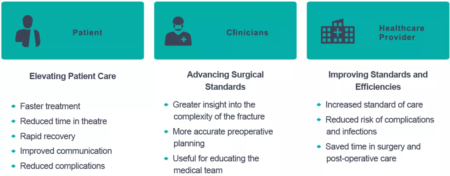
Using the 3D printed models, trial operations can be carried out to improve planning and reduce the surgical time associated with difficult cases and allow much easier communication with patients and clinicians. Articular lesions and deformity are particularly clear on the models, compared to standard images.
Benefits
Do you have a complicated patient case? What could we 3D print for your speciality? Find out if you’re eligible for a free patient-specific 3D printed model of your next patient case.
Request a free anatomical model
Case Study8th November 2023
Enhancing Surgical Precision: Patient-Specific 3D Model for Vascular Calcification Assessment
Case Study28th August 2023
Optimizing surgical outcomes with 3D models in Double Outlet Right Ventricle (DORV) cases
Case Study28th August 2023
Gaining insights into anatomical intricacies of ccTGA using patient-specific 3D solutions
Case Study2nd December 2021
Correcting Transposition of the Great Arteries with the Help of a Patient-Specific 3D Printed Anatomical Model
Case Study29th November 2021
Medical 3D model saves crucial time in heart transplant surgery for a patient with congenital heart disease
Follow us


