Case Study18th January 2024
Revolutionizing Pediatric Cardiac Care at Southampton Hospital
3D Printed Model Enhances Pre-Operative Planning for Complex Calcaneal Fracture
Case Study
A male patient was admitted to Ulster Hospital with severe injuries to his left calcaneus due to a crushing incident. The intricate and closely positioned anatomical structures within the foot made it challenging to obtain a comprehensive assessment of the extent of the injury using conventional CT scans.
Solution
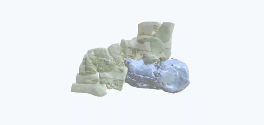
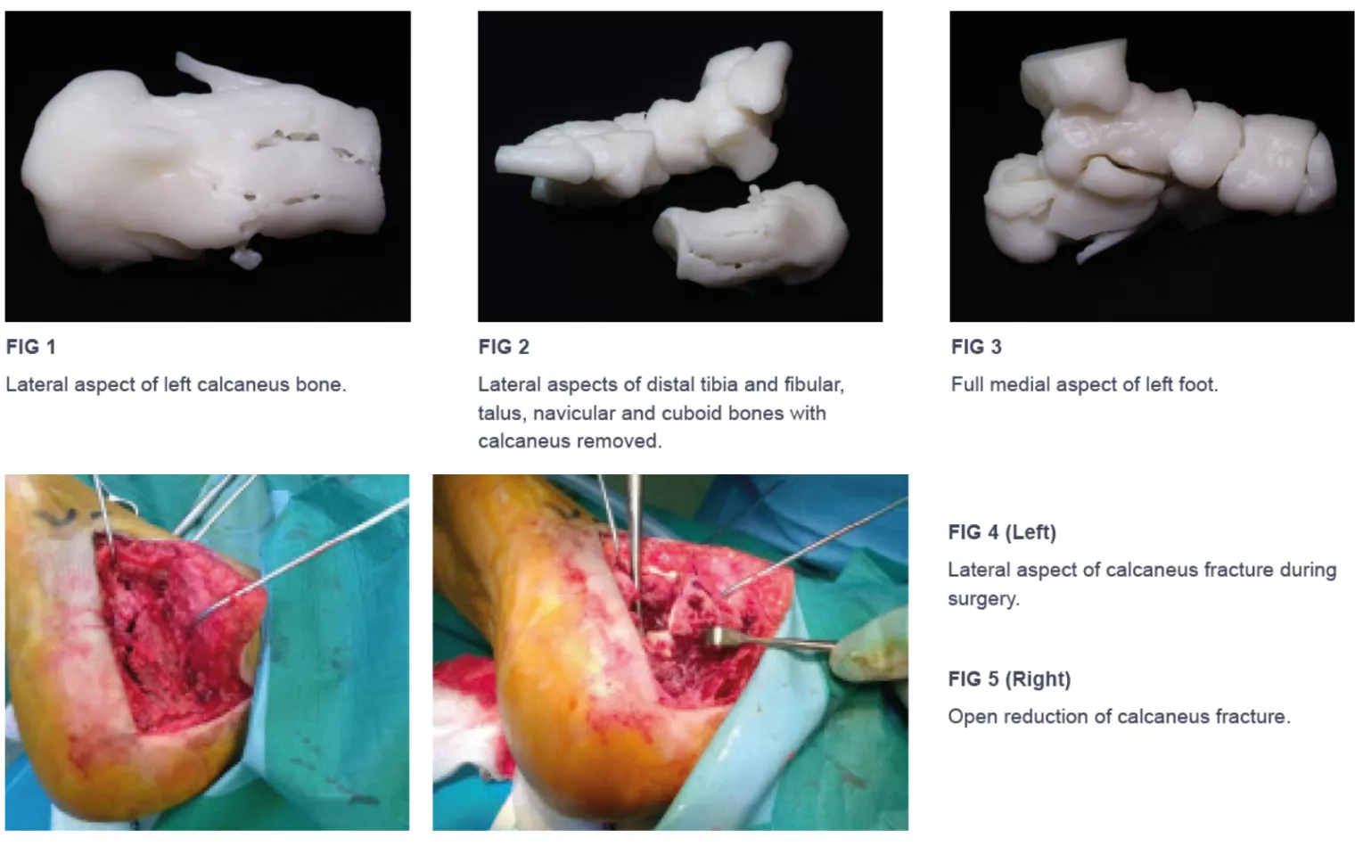
The surgical team turned to Axial3D to address this complex case. They requested a 1:1 scale 3D-printed model of the calcaneal fracture, intricately detailing the surrounding foot anatomy, including the distal aspects of tibia and fibula, talus, navicular, and cuboid bones (Figure 1, 2, and 3).
Results
The 3D printed model proved invaluable in gaining a deeper understanding of the fracture's complexity and enabled precise planning for open reduction and internal fixation (Figure 4 and 5). Furthermore, the model served as a powerful educational tool, allowing medical staff to grasp the extent of the patient's injuries before surgery.
"I was provided with a 3D physical model of a complex calcaneal fracture, which allowed me to understand the complexity of the fracture and plan open reduction and internal fixation. The model was of a high standard and was extremely useful in pre-operative planning and educating medical staff."
Mr. A. Adair, Consultant Orthopedic Surgeon, Ulster Hospital, Northern Ireland
Conclusion
Leveraging 3D printed models proves to be a game-changer in complex surgical cases, enhancing pre-operative planning and reducing surgical duration. These models offer unparalleled clarity in visualizing articular sites and fractures, surpassing conventional imaging techniques. Moreover, they facilitate improved communication with patients and post-operative healthcare personnel, ushering in a new era of precision and collaboration in orthopedic surgery.
Talk to our experts about your next upcoming case.
Contact us.
Case Study8th November 2023
Enhancing Surgical Precision: Patient-Specific 3D Model for Vascular Calcification Assessment
Case Study28th August 2023
Optimizing surgical outcomes with 3D models in Double Outlet Right Ventricle (DORV) cases
Case Study28th August 2023
Gaining insights into anatomical intricacies of ccTGA using patient-specific 3D solutions
Case Study2nd December 2021
Correcting Transposition of the Great Arteries with the Help of a Patient-Specific 3D Printed Anatomical Model
Case Study29th November 2021
Medical 3D model saves crucial time in heart transplant surgery for a patient with congenital heart disease
Follow us


