Case Study18th January 2024
Revolutionizing Pediatric Cardiac Care at Southampton Hospital
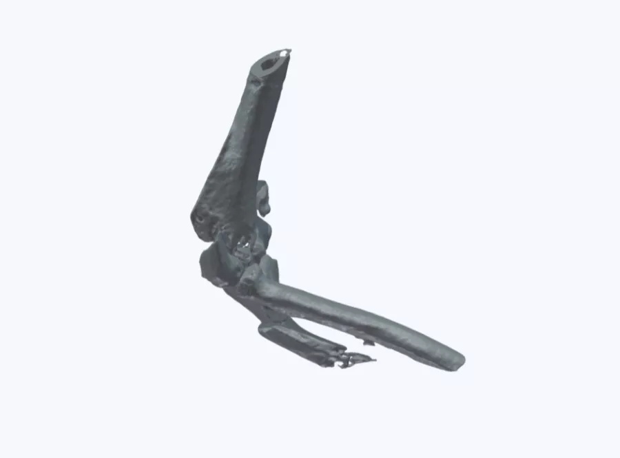
Fracture surgery planning transformed with 3D modeling
Case Study
The medical 3D model used for this case resulted in the following benefits:
- Improved patient consent process
- Saved 40 minutes of pre-operative planning time
- Enabled more accurate (and less needed) surgery equipment selection
Case
This 36-year-old man presented with a severe fracture in his upper arm, causing pain, swelling and discomfort. Most fractures of this type can be treated without surgery, but unfortunately in this case the patient had to undergo surgery as the bone fragments were so badly displaced.
Solution
Due to the complex nature of the injury, a 3D model was requested to help the team get a closer look at the problem and to help communicate the plan to the patient. The model supported better communication between the team as they defined their plan and reassured them that they had selected the safest and best route for the patient. With the model, the team managed to confirm this procedure plan 40 minutes faster than they would have done using 2D medical scans alone.
This type of surgery will usually involve fixation of the fractured fragments with plates, screws or pins, which are often selected intraoperatively.
Benefits of the 3D Model
The 3D model enabled the team to take a more targeted approach in selecting the exact equipment that they needed ahead of time, reducing equipment costs, waste and ultimately saved a further 20 minutes while in the OR and ensuring a positive outcome for the patient.
Experience Axial3D for yourself
Request a free anatomical model
Case Study8th November 2023
Enhancing Surgical Precision: Patient-Specific 3D Model for Vascular Calcification Assessment
Case Study28th August 2023
Optimizing surgical outcomes with 3D models in Double Outlet Right Ventricle (DORV) cases
Case Study28th August 2023
Gaining insights into anatomical intricacies of ccTGA using patient-specific 3D solutions
Case Study2nd December 2021
Correcting Transposition of the Great Arteries with the Help of a Patient-Specific 3D Printed Anatomical Model
Case Study29th November 2021
Medical 3D model saves crucial time in heart transplant surgery for a patient with congenital heart disease
Follow us


