Case Study18th January 2024
Revolutionizing Pediatric Cardiac Care at Southampton Hospital
Intracranial aneurysm surgery aided by 3D printed model
Case Study
Case
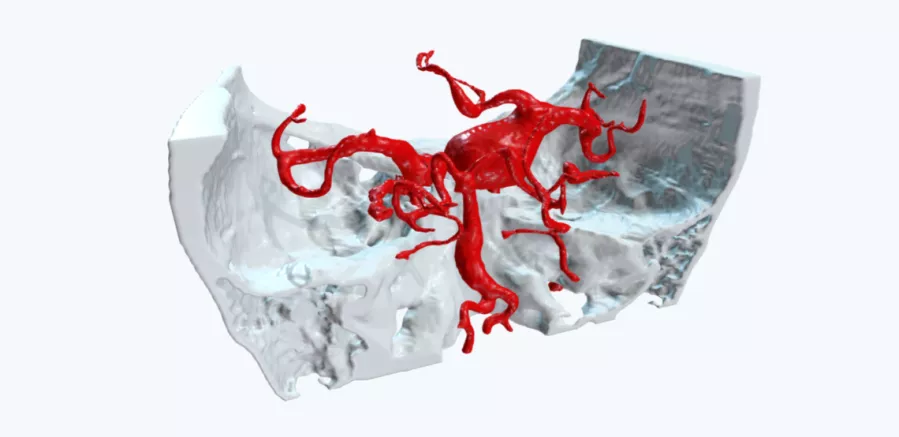
A 77-year-old female presented with a large basilar arterial aneurysm. The CT images used made it difficult to conceptualize the size of the vessel and the proximity of the vessel to other surrounding arteries.
The CT images were used to create a 1:1 scale flexible model of the aneurysm and surrounding anatomy. The model was used to plan the exact trajectory of the catheter stent deployment. The stent was also measured against the 3D printed model prior to surgery.
Outcomes
The clinicians had an improved diagnosis of the intracranial aneurysm and used the model to create an exact plan ahead of surgery. This resulted in a substantial reduction of risk for misplacement or wrong stent being used, drastically improving outcomes for the patient.
Try a 3D printed anatomical model for an upcoming case.
Request a free anatomical visual
Case Study8th November 2023
Enhancing Surgical Precision: Patient-Specific 3D Model for Vascular Calcification Assessment
Case Study28th August 2023
Optimizing surgical outcomes with 3D models in Double Outlet Right Ventricle (DORV) cases
Case Study28th August 2023
Gaining insights into anatomical intricacies of ccTGA using patient-specific 3D solutions
Case Study2nd December 2021
Correcting Transposition of the Great Arteries with the Help of a Patient-Specific 3D Printed Anatomical Model
Case Study29th November 2021
Medical 3D model saves crucial time in heart transplant surgery for a patient with congenital heart disease
Follow us


