Case Study18th January 2024
Revolutionizing Pediatric Cardiac Care at Southampton Hospital
Sixty minutes of surgery time saved in neurotrauma case following car accident
Case Study
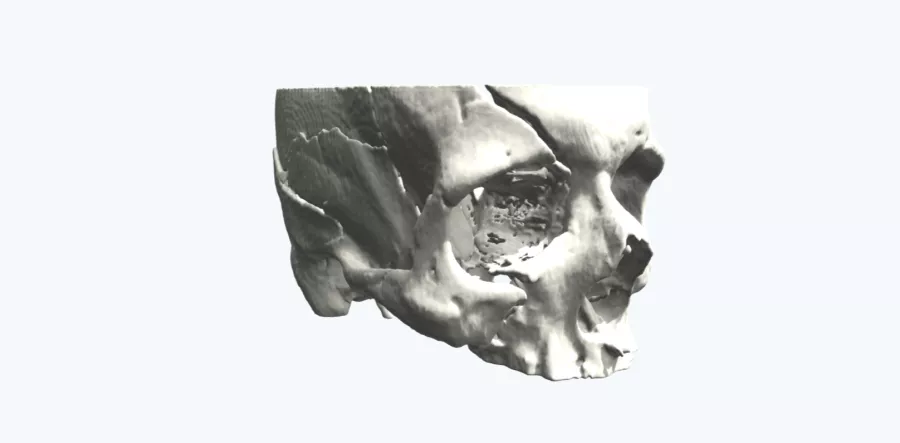
A 27-year-old male presented with extensive facial trauma following a road traffic collision. The fracture plane crossed multiple portions of anatomy from the maxilla superiorly to the frontal bone. Given the extensive fracture caused by the accident, treatment planning was considered difficult utilizing the 2D CT imagery alone. In addition, the case required collaboration and planning from both neurological and cranio-maxillofacial teams.
Case
A full-scale model was created from the patient’s CT scans. The surgeons used the 3D printed model to choose, measure and prepare all the required equipment for surgery. The model was utilized within a multidisciplinary planning meeting to mutually conclude on the best surgical approach.
Outcome
Sixty minutes of surgical time was saved as a result of using the 3D model in the planning. This significant reduction in operative time helped to elevate the patient's time in care in the form of faster treatment, reduced OR time with less invasive surgery, a more rapid recovery and reduced complications. Only 2 surgical trays needed to be sterilized, reduced from 6, as a result of choosing equipment prior to surgery, adding to the hard cost savings realized by the hospital.
Up next: 3D printed aneurysm model used to practice procedure and pre-select equipment before surgery
Request a free personalized 3D model Axial3D makes it easy for you to access patient-specific 3D printed models. We create and ship a 3D printed model from your 2D CT or MRI images within 48 hours – allowing you to concentrate on what matters most, your patients.
Request a free anatomical visual
Case Study8th November 2023
Enhancing Surgical Precision: Patient-Specific 3D Model for Vascular Calcification Assessment
Case Study28th August 2023
Optimizing surgical outcomes with 3D models in Double Outlet Right Ventricle (DORV) cases
Case Study28th August 2023
Gaining insights into anatomical intricacies of ccTGA using patient-specific 3D solutions
Case Study2nd December 2021
Correcting Transposition of the Great Arteries with the Help of a Patient-Specific 3D Printed Anatomical Model
Case Study29th November 2021
Medical 3D model saves crucial time in heart transplant surgery for a patient with congenital heart disease
Follow us


