Case Study18th January 2024
Revolutionizing Pediatric Cardiac Care at Southampton Hospital
Providing unparalleled insights and a significant reduction in procedure time with 3D printing
Case Study
The Case
The patient presented with renal failure, requiring an urgent transplant, however, to add to the complications, he was also suffering from a severe case of hyperparathyroidism - an enlargement of one of the parathyroid glands which cause hypercalcemia (high levels of calcium in the blood). This resulted in the over-calcifying and hardening of vessels in the patient.
If the kidney transplant were to be carried out with the parathyroid still overproducing calcium, the kidney would be rejected, resulting in renal failure, and more suffering for the patient - a recipe for a serious life-threatening situation.
Ahead of the patient becoming eligible for transplant, the surgeons made a plan for the parathyroid gland to be removed to bring the calcium levels back to a normal level.
Over-activity of the parathyroid glands can lead to higher than usual calcium levels in the blood, which left untreated, can lead to osteoporosis and bone pain, muscle weakness, kidney stones, and other complications.
The Solution
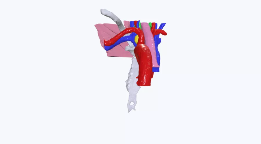
In this case, the gland was severely enlarged and was situated between 2 vital vascular structures, the aorta, and vena cava. It also had a complex array of anatomy overlaid on top, meaning the trajectory to the gland was extremely difficult to visualize. Because of the location of the gland, two surgeries to remove it were unsuccessful as the surgeons were unable to locate the gland safely for removal. As these anatomical structures are located in a small space within the patient’s neck, clinicians rely on technology to provide them with pinpoint accuracy and detailed levels of insight prior to operating.
Until the hypercalcemia was brought under control, the patient was unable to qualify for a kidney transplant. The timely removal of the gland proved critical to the patient’s ability to receive a donor's kidney.
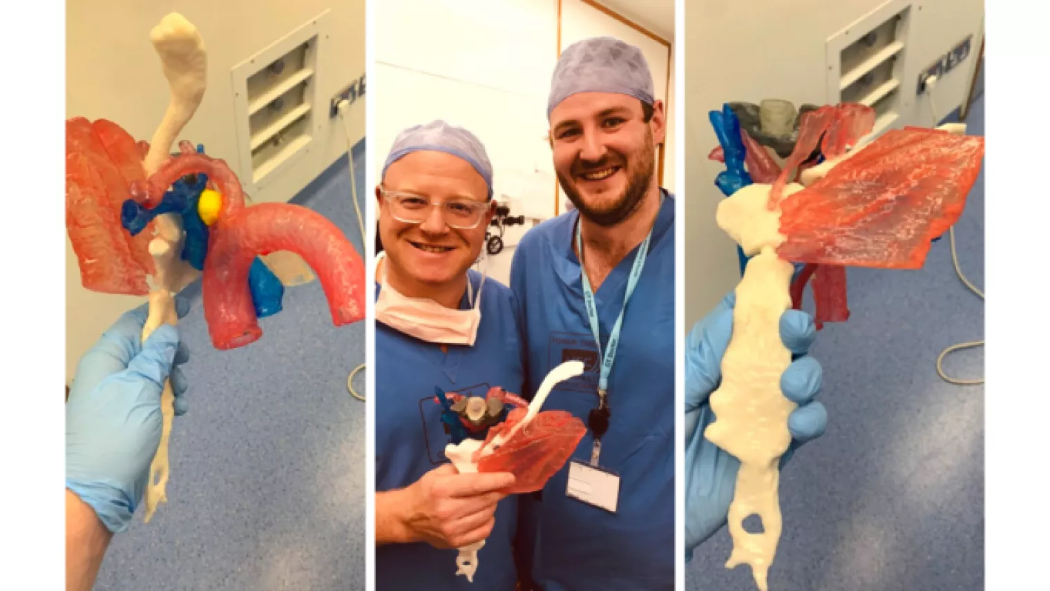
Prior to conducting a third procedure to remove the gland, a 3D printed model of the patient’s anatomy was requested in order to give the insight required to enable the clinical team to safely locate and excise the gland. The model, printed at a 1:1 scale, proved invaluable to the team in planning for the third surgery and in gaining consent from the patient to proceed. At the request of the surgeon, the enlarged parathyroid gland was contrasted in yellow to help it stand out against the rest of the anatomy, while the scapula was printed in white standard resin, mimicking bony anatomy.
Benefits of using the 3D Model
In this case, having access to the 3D printed model enabled:
- A significant reduction in time and cost of the procedure
- Enhanced pre-operative planning
- Surgical simulation
- Intrateam discussions
- Patient consent discussions
Mr. Tim Brown, consultant transplant surgeon at Belfast City Hospital, said, “When it comes to ordering a 3D model from Axial3D, there is no heavy lifting at all. I take anonymized patient data and I send it through to Axial3D with a couple of clicks of a button. A few days later, there's a delivery with a beautiful life-size, 3D representation model, which I can then take to plan and treat a patient with. From my perspective, it's as easy as sending an email, which we do hundreds of times a day. ”
Having access to the 3D printed model, the third attempt to locate and excise the enlarged parathyroid gland was a success. The insight provided by the model in the planning stages enabled the clinical team to safely remove the gland in just 20 minutes. A standard parathyroid operation can typically take 3.5 to 4 hours but may take as long as 8 hours. In this case, having the 3D model reduced this timeframe significantly and aided a successful procedure.
The patient’s calcium levels dropped significantly overnight and he successfully underwent a kidney transplant just 8 days later. The patient now has a healthy, functioning kidney & full life expectancy.
Do you have a complicated patient case? What could we 3D print for your speciality? Find out if you’re eligible for a free patient-specific 3D printed model of your next patient case.
Request a free anatomical visual
Case Study8th November 2023
Enhancing Surgical Precision: Patient-Specific 3D Model for Vascular Calcification Assessment
Case Study28th August 2023
Optimizing surgical outcomes with 3D models in Double Outlet Right Ventricle (DORV) cases
Case Study28th August 2023
Gaining insights into anatomical intricacies of ccTGA using patient-specific 3D solutions
Case Study2nd December 2021
Correcting Transposition of the Great Arteries with the Help of a Patient-Specific 3D Printed Anatomical Model
Case Study29th November 2021
Medical 3D model saves crucial time in heart transplant surgery for a patient with congenital heart disease
Follow us


