Case Study18th January 2024
Revolutionizing Pediatric Cardiac Care at Southampton Hospital
Simulating a surgical plan with physical 3D anatomical models
Case Study
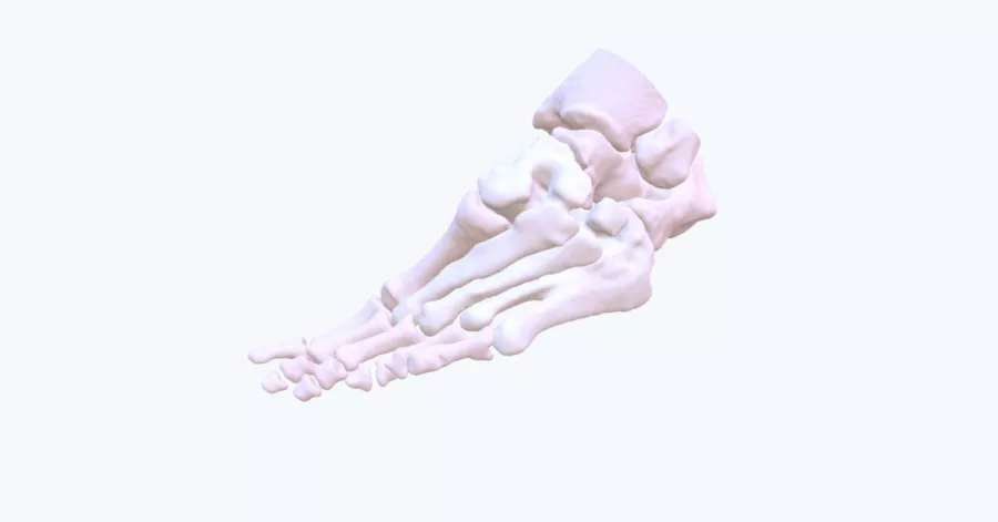
A patient presented with a severe deformity affecting the majority of the bones in their foot, most notably in the 3rd, 4th, and 5th metatarsal. The surgeon found it difficult to fully conceptualize the deformity on 2D images given the complex arrangements of the bones.
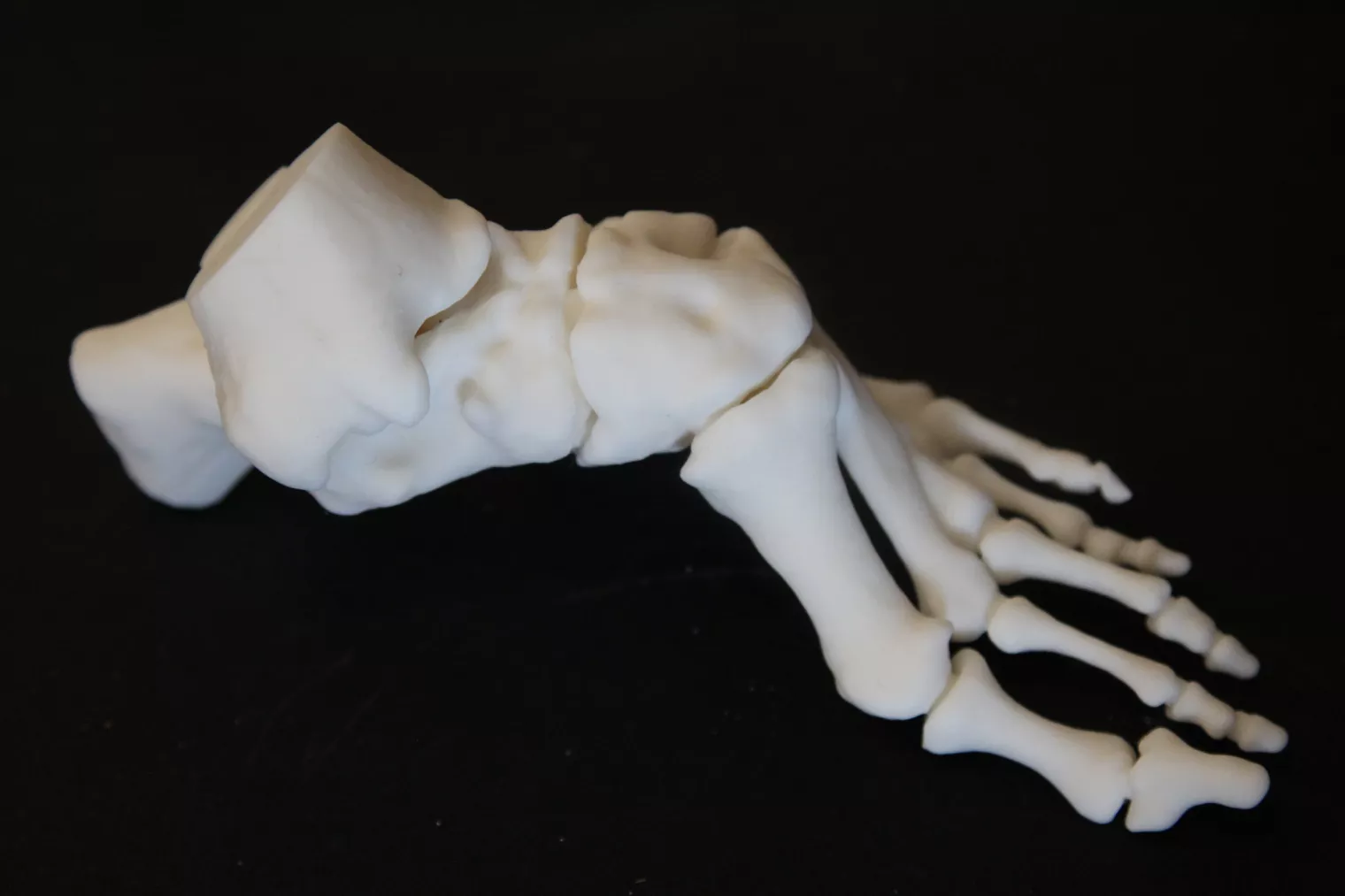
A 3D printed model was requested by the surgeon as a pre-operative planning guide to help him gain greater insight into the entire pathology.
Benefits of the 3D Model
“The model enabled us to gain a much greater insight into the patient's entire deformity. We were also able to place the physical models of the patient's foot onto flat surfaces to plan how we were going to correct their gait with equipment prior to surgery. This greatly improved our confidence of a successful surgery and was used to explain the procedure to clinical staff and registrars.”
– Mr. John Wong, Consultant Orthopedic Surgeon, Altnagelvin Hospital
The model was also used to practice several different osteotomy approaches to ensure the correct plan and equipment was prepared before stepping into the theatre.
Without access to the 3D model, the surgeon would not have had the opportunity to physically simulate his plan to correct the patient’s gait, nor would he have been able to select the exact equipment ahead of time, resulting in longer operating times and costs.
Try Axial3D with a free anatomical model of one of your cases
Request a free anatomical visual
Case Study8th November 2023
Enhancing Surgical Precision: Patient-Specific 3D Model for Vascular Calcification Assessment
Case Study28th August 2023
Optimizing surgical outcomes with 3D models in Double Outlet Right Ventricle (DORV) cases
Case Study28th August 2023
Gaining insights into anatomical intricacies of ccTGA using patient-specific 3D solutions
Case Study2nd December 2021
Correcting Transposition of the Great Arteries with the Help of a Patient-Specific 3D Printed Anatomical Model
Case Study29th November 2021
Medical 3D model saves crucial time in heart transplant surgery for a patient with congenital heart disease
Follow us


