Case Study18th January 2024
Revolutionizing Pediatric Cardiac Care at Southampton Hospital
Enhancing Precision in Orthopaedic Trauma: A Case Study on Comminuted Both Column Acetabular Fracture
Case Study
Case
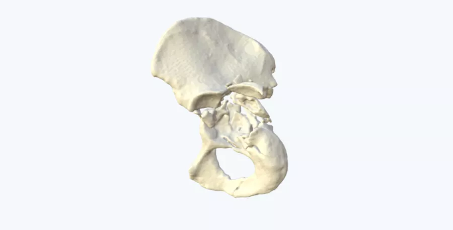
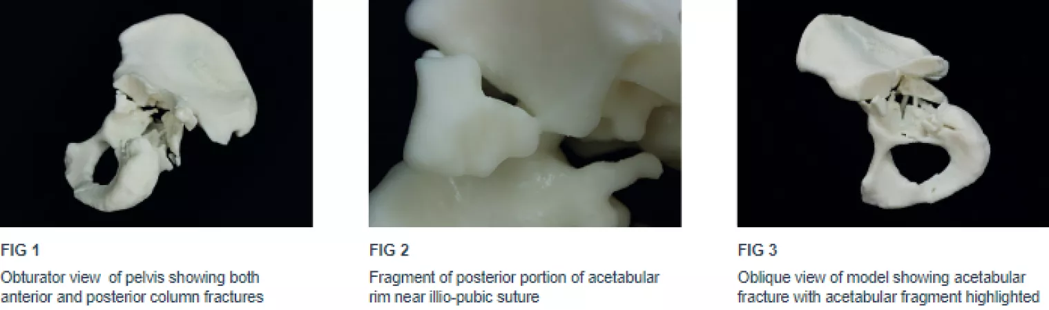
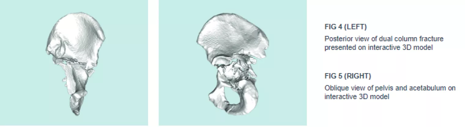
A patient presented with severe trauma following a motorcycle accident which resulted in an associated both column fracture of the acetabulum. Due to the severity and comminution of the fracture, 2D imaging did not convey the full extent of the fracture pattern and fragment location. This made it difficult to conceptualize the pathology. The severity of the injury necessitated a higher level of insight.
Solution
A physical, 1:1 scale 3D printed model of the patient’s pelvis provided additional insight into the severe injury beyond what was possible by viewing traditional 2D patient scans alone. The model helped the surgical team determine the optimum treatment plan.
Result
With access to the 3D printed model the clinical team came to the consensus that the original plan was to change from a posterior approach to a dual anterior and posterior approach. This was concluded following the difficulty in visualizing the entirety of the fracture pattern, including fragment location that was not seen from the 2D images.
Conclusion
The model allowed the trauma team to gain a much greater insight into the patient’s pathology. With access to the 3D printed model, the optimum surgical treatment was determined; significantly reducing the surgery time and improving patient outcome following this severe fracture.
Do you have a complicated patient case?Let us help. Find out if you're eligible for a free 3D visual today.
Request a free anatomical visual
Case Study8th November 2023
Enhancing Surgical Precision: Patient-Specific 3D Model for Vascular Calcification Assessment
Case Study28th August 2023
Optimizing surgical outcomes with 3D models in Double Outlet Right Ventricle (DORV) cases
Case Study28th August 2023
Gaining insights into anatomical intricacies of ccTGA using patient-specific 3D solutions
Case Study2nd December 2021
Correcting Transposition of the Great Arteries with the Help of a Patient-Specific 3D Printed Anatomical Model
Case Study29th November 2021
Medical 3D model saves crucial time in heart transplant surgery for a patient with congenital heart disease
Follow us


