Case Study18th January 2024
Revolutionizing Pediatric Cardiac Care at Southampton Hospital
When 2D imaging doesn't cut it: 3D modeling for enhanced insights
Case Study
Case
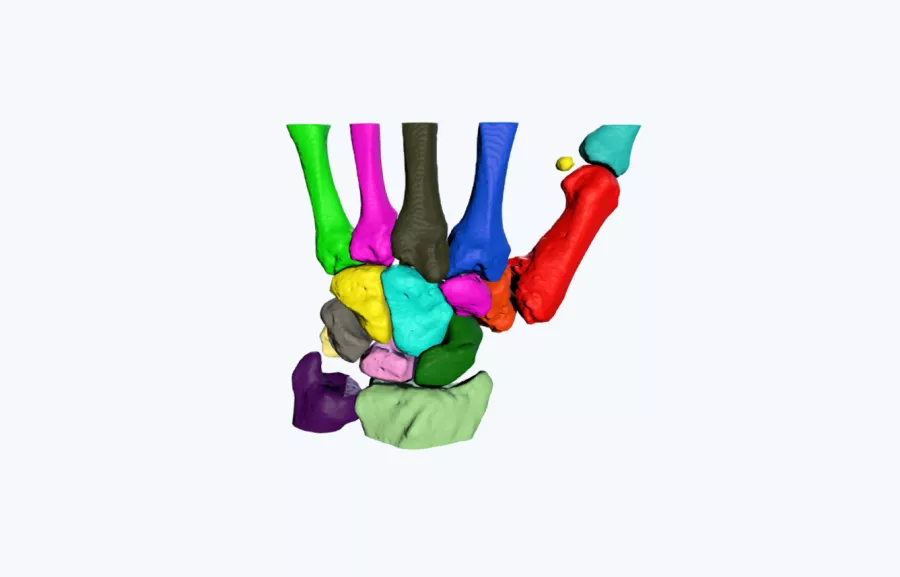
A 3D printed model was used to improve the understanding of the size and morphology of a deformed trapezium. Due to the complex anatomical arrangement of the carpal bones with the articulating aspects of the metacarpals, the understating of the anatomy of the bone was extremely difficult to conceptualize on CT scans alone.
The 3D printed anatomical model enabled the surgeon to gain a much greater insight into the shape of the bone, which improved his confidence that surgery was indeed the correct approach.
Outcome
“Having the 3D printed model allowed me to see the whole deformity on the trapezium which was not possible with the original CT scans provided. The pathology was much more complex than initially diagnosed, confirming my decision-making that surgical intervention was required.”
– Mr Jeremy Field, Consultant Orthopaedic and Hand Surgeon, Cheltenham General Hospital, Gloucester
Do you have a complicated patient case? Get a free patient-specific 3D printed model of your next patient case.
Request a free anatomical visual
Case Study8th November 2023
Enhancing Surgical Precision: Patient-Specific 3D Model for Vascular Calcification Assessment
Case Study28th August 2023
Optimizing surgical outcomes with 3D models in Double Outlet Right Ventricle (DORV) cases
Case Study28th August 2023
Gaining insights into anatomical intricacies of ccTGA using patient-specific 3D solutions
Case Study2nd December 2021
Correcting Transposition of the Great Arteries with the Help of a Patient-Specific 3D Printed Anatomical Model
Case Study29th November 2021
Medical 3D model saves crucial time in heart transplant surgery for a patient with congenital heart disease
Follow us


